In the world’s first, London surgeons have used real-time AI to protect a patient's sight during live brain surgery. See what the trial’s AI framework means for MedTech.
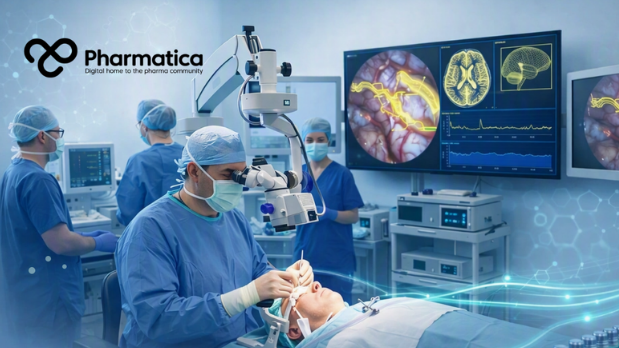
Surgeons in London have carried out the world’s first live AI-assisted brain surgery, using real-time computer vision to protect a patient’s eyesight during a delicate pituitary tumour operation.
Rhys Hibbert, a 48-year-old customer services manager from Bedfordshire, is the first patient to undergo surgery this way. His pituitary tumour was found by chance in December 2024, after he collapsed and had a seizure during a walk.
Left untreated, the tumour would have continued pressing on the structures that control vision. Doctors warned it could ultimately have caused blindness.
Professor Hani Marcus, a consultant neurosurgeon, performed the procedure alongside Danyal Khan, a UCL PhD candidate and neurosurgical resident who leads the technical work.
During the operation, an AI system analysed the live endoscopic video feed and highlighted critical anatomy at the base of the brain in real time.
In this cramped space, the pituitary gland, major blood vessels, and the nerves controlling sight sit only millimetres apart.
With the help of AI, the surgery successfully removed the tumour and protected Hibbert’s vision. To the Guardian newspaper, he described seeing “everything in the room clearly” upon waking, and was walking independently within a week, without the glasses or walking sticks he had relied on before.
How the AI System Sees Inside the Brain in Real Time
To operate effectively within complex neurosurgical environments, real-time surgical assistance requires advanced imaging architectures and high-performance processing hardware that go beyond traditional pre-operative static planning.
Reading live video, not pre-operative scans
Most existing surgical navigation tools rely on probes or static scans taken before the operation begins. However, anatomy shifts once surgery is under way, so pre-operative images can quickly become unreliable guides.
The UCL system instead works from the live surgical video itself. It was built to flag risky areas for the surgeon to avoid, while still allowing as much of the tumour to be safely removed as possible, and it has the potential to track surgical instruments and how they interact with tissue.
An edge-computing platform built for the operating room
The system runs on an NVIDIA Clara IGX platform and uses a DINOv3-derived vision transformer, a type of computer vision model. It was developed at the UCL Hawkes Institute, with Dr Sophia Bano, Associate Professor in Robotics and Artificial Intelligence, as technical lead.
Crucially, AI inference happens on-site, on dedicated hardware, rather than in the cloud. That design choice matters for a procedure where a delayed response is not an acceptable trade-off.
Researchers trained the model on a large library of annotated endoscopic pituitary surgery videos gathered from previous operations.
A Trial Built on Evidence Frameworks, Not Just Ambition
The operation is part of a formally registered clinical trial (NCT07568366) and follows two established evaluation frameworks for early-stage device innovation: IDEAL and DECIDE-AI.
Both frameworks exist to stop promising technology reaching patients before its risks are properly understood.
This first stage, described in a preprint from the research team, was a small proof-of-concept study rather than a large efficacy trial. Six patients with pituitary adenomas were enrolled, and the results were mixed but instructive:
The AI system was successfully deployed in four of the six cases, with acceptable accuracy in outlining the sella, the bony structure housing the pituitary gland.
Deployment failed before surgery in the remaining two cases, due to a recurring system reboot bug.
Between cases, the team added new anatomical structures for the AI to identify, including the carotid arteries, and pushed through hardware and model updates.
Both live observation and blinded video review found no adverse events and no meaningful distraction to the lead surgeon.
Surveys of the surgical team reported satisfactory feasibility, usability, and acceptability.
Notably, the system was deployed as an educational adjunct on a secondary monitor for this first stage, so the primary surgical feed was never compromised.
Direct, in-line decision support is described as a future step, not the current reality.
“This is one of the riskiest industries there is because you're dealing with human lives, you're dealing with experimental protocols, and you're dealing with regulatory bodies where you might not get another shot at that clinical trial."
Image
What the Trial Design Signals for MedTech
When evaluating AI-enabled devices, the choice of framework matters as much as the headline result.
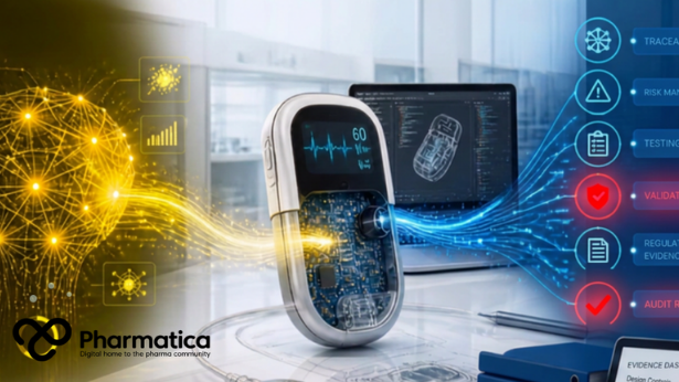
The IDEAL and DECIDE-AI frameworks ensure developers document failure modes, iterate transparently between cases, and report human factors data alongside clinical outcomes, rather than moving straight to a large trial on the strength of a promising demo.
That staged, failure-tolerant approach is noteworthy for any teams developing AI-driven decision support alongside a drug, device, or diagnostic.
Regulators are increasingly likely to expect this kind of evidence trail for software as a medical device.
The funding and governance picture is equally instructive. The trial was backed by the NIHR and Google, alongside the Royal College of Surgeons, the Engineering and Physical Sciences Research Council, and Wellcome.
The published preprint also discloses that two of the senior investigators hold shares in Panda Surgical, a spin-out company, and have other industry ties.
That transparency is a useful reference point. As more clinicians co-develop the AI tools they later evaluate, disclosed equity and consulting relationships will draw closer scrutiny from ethics boards, journals, and investors alike.
This blended funding structure, spanning a public research funder, a technology company, and a medical research charity, is likely to become more common as surgical AI moves from academic prototype toward commercial product.
Pharma and MedTech scoping their own partnerships should note how clearly this trial separated the roles of funder, developer, and clinical evaluator, even while some individuals held more than one of those roles at once.
The Road from Proof-of-Concept to Everyday Practice
This proof-of-concept stage is only the beginning. The research team has already outlined next steps, moving toward a larger single-centre case series, known as IDEAL Stage 2a, involving more surgical teams.
Several milestones will need to be cleared before real-time surgical AI becomes routine, notably
Validation across multiple centres and surgical teams, not just the unit that built the system.
A regulatory pathway suited to software that updates iteratively between cases, rather than shipping as a fixed product.
Integration with existing intra-operative navigation technology and hospital IT infrastructure.
A credible reimbursement model, since edge-computing hardware and ongoing model maintenance carry real costs.
Continued human factors research, so surgeons trust the system's confidence signals rather than treating it as a black box.
Other research groups are exploring adjacent applications, including AI that highlights tumour tissue on intra-operative ultrasound, or flags the approximate location of critical structures.
Across neuroscience, the move is from static, pre-operative planning tools towards live, adaptive support inside the theatre itself.
For teams with neuro-oncology or ophthalmology portfolios, this trajectory must be watched closely. A surgical AI system that can reliably map critical anatomy in real time could eventually inform how surgical resection margins are defined, which, in turn, could influence how adjuvant drug therapies are sequenced and timed after surgery.
Image
Five Questions Pharma Leaders Are Asking about AI-Assisted Neurosurgery
The world’s first successful AI-assisted neurosurgery has now been performed and has helped preserve the vision of a patient. Its case study raises important questions for MedTech and pharma innovators.
What is AI-assisted brain surgery?
This might feel and sound like a stupid and too basic question, but the answer is worth repeating!
AI-assisted brain surgery uses computer vision software to analyse a live surgical video feed during an operation, highlighting critical anatomy such as blood vessels and nerves in real time.
It is designed to support the surgeon's judgement, not to make decisions independently.
How does the UCL and UCLH AI system work during surgery?
The system uses a vision transformer model, run on dedicated edge-computing hardware in the operating room, to interpret the endoscopic video feed as surgery happens.
It was trained on hundreds of annotated recordings of previous pituitary operations and currently runs on a secondary monitor as an educational adjunct.
Is AI-assisted neurosurgery safe?
In this first-in-human evaluation, researchers reported no adverse events linked to the AI system and no meaningful distraction to the lead surgeon, based on both live observation and blinded video review.
The evidence base is still small and growing, drawn from six patients, and larger studies are planned to confirm safety at scale.
What regulatory pathway applies to real-time surgical AI?
This trial followed the IDEAL and DECIDE-AI frameworks with staged approaches used to evaluate surgical innovations and AI-enabled devices before wider rollout.
These frameworks are increasingly referenced as a template for how regulators may expect real-time clinical AI to be evidenced.
What does this mean for pharma and MedTech investment in surgical AI?
The trial shows that rigorous, transparent, staged evaluation, including disclosed funding sources and researcher financial interests, can coexist with fast-moving AI development.
Companies developing AI-enabled devices or companion decision-support tools can look to this framework when planning their own clinical evidence strategy.
AI in Pharma: Why the Future of Healthcare Starts With Patients, Not Tech
Kate O’Reilly, President & Chair of the Healthcare Businesswomen’s Association (HBA) Dublin-Ireland Chapter & Healthcare Transformation Partner at Roche, discusses AI in pharma, patient engagement, and the future of healthcare innovation.
Image
Connecting Innovation, Evidence, and Surgical Practice
The world’s first live AI-assisted brain surgery demonstrates how real-time computer vision can actively assist surgeons in protecting critical anatomical structures during complex procedures.
Beyond the headline clinical success, the trial’s reliance on structured evidence frameworks like IDEAL and DECIDE-AI provides a clear blueprint for how AI-enabled medical devices can demonstrate safety, navigate regulatory pathways, and build clinical trust.
As surgical AI transitions from early proof-of-concept studies toward broader clinical practice, establishing transparent governance, edge-computing infrastructure, and multi-centre validation will be essential to unlocking its full therapeutic impact.
Pharmatica will continue reporting the IDEAL Stage 2a results as the trial expands to more surgical teams. For more analysis on how AI-enabled devices are reshaping clinical practice, explore our Digital Pulse coverage, or get in touch with our editorial team to discuss your organisation’s own evidence strategy.
Pharmatica: Insight. Connection. Impact.
Frequently Asked Questions
What is AI-assisted brain surgery?
AI-assisted brain surgery uses real-time computer vision models to analyse live endoscopic video feeds during an operation. The software identifies critical anatomical structures (such as blood vessels, nerves, and glands) to assist the surgeon's navigation and decision-making without operating independently.
How does real-time surgical AI differ from traditional surgical navigation tools?
Traditional surgical navigation tools rely on pre-operative static scans, which can become inaccurate as anatomical structures shift during surgery. Real-time surgical AI processes live intra-operative video directly, allowing it to adapt continuously to anatomical changes during the procedure.
What role do the IDEAL and DECIDE-AI frameworks play in AI medical device trials?
The IDEAL and DECIDE-AI frameworks provide structured, staged evaluation guidelines specifically designed for surgical innovations and early-stage clinical AI decision-support systems. They ensure developers evaluate human factors, document software failure modes, and gather safety and usability data before advancing to large-scale clinical trials.
Why is edge computing important for AI in the operating room?
Edge computing allows AI algorithms to perform inference locally on dedicated hardware within the operating room rather than sending data to cloud servers. This eliminates network latency, ensuring real-time processing speeds essential for live surgical feedback.
How can pharma and MedTech companies leverage surgical AI clinical trials?
Surgical AI can help define precise resection margins, potentially influencing post-operative treatment strategies and adjuvant drug therapy sequencing. Additionally, following established evidence frameworks helps life science companies build transparent, regulator-approved clinical evidence strategies for novel digital health tools.
Nicole (BSc Molecular Medicine, Honours Medical Biochemistry) has many years of pharmaceutical experience, having worked for top CROs and biopharma companies for more than a decade.
Did you enjoy the content?
3
Why not support
Nicole Dale
by giving this content a like
AI Creates New Viruses? Not Quite. AI-Designed Bacteriophages
AI-designed bacteriophages show how genome models could create new antibacterial therapies while forcing pharma to rethink biological AI governance and biosecurity.
Corpus Healthy Human Protein Atlas Gives AI a Better Map
The healthy human protein atlas from Ambitious Bio could sharpen target safety checks in AI drug discovery. Explore the claims, the gaps, and the questions to ask.
Ketryx and Accenture Build Regulated AI MedTech at Scale
Ketryx and Accenture are combining AI-native compliance infrastructure and transformation expertise to help MedTech scale regulated AI without slowing development.
AI Breast Cancer Screening Moves From Trials to Policy
AI breast cancer screening finds more cases and cuts radiologist workload, but recall risks remain. See what MASAI, AITIC, and EDITH mean for MedTech teams.
How AI Is Reshaping Data Integrity in Pharma Manufacturing
Veragence CEO Nour AlSaleh discusses how AI is transforming data integrity in pharma manufacturing, regulatory compliance, the "human-in-the-loop" requirement, and patient safety in the era of AI-driven GxP systems.











Comments (0)